Abstract:
Narcolepsy is a disorder caused by losing orexin neurons, marked by excessive daytime sleepiness and cataplexy, sudden weakening of muscle tone. Previously, we found two kinds of neurons preventing narcolepsy by receiving information from orexin neurons. Here, we have discovered that serotonin neuron, one of the two neurons above, inhibits cataplexy by reducing the amygdala activity, which controls emotion. Current discovery should lead to comprehension of the narcolepsy mechanism and to therapy development for cataplexy.
[Background]
Sleep is of absolute necessity for us humans, although if one falls asleep all of a sudden while being awoken, it would cause a big trouble. The brain is equipped with sleep mechanism and wakefulness mechanism, which are regulated to be on or off in an adequate manner. It is orexin*1 that is important in regulating this switch. If orexin neurons are lost, one suffers from narcolepsy*2, a sleep disorder, where sleep and wakefulness are inadequately switched on and off. The typical symptoms are excessive daytime sleepiness and cataplexy*3. Cataplexy takes place when one is very excited in terms of one’s emotion and if severe, one may lose the muscle tone of the whole body and fall down.
Sleep is categorized into two, REM sleep and non-REM sleep. Dreams are dreamt usually during REM sleep, where most of the muscles are controlled to be relaxed (called atonia) in order to prevent the dreamer to make real actions. Cataplexy attack is thought that atonia, a characteristics of REM sleep, takes place while one is awoken. The research team previously found two types of neurons preventing narcolepsy by receiving orexin from orexin neurons. The one is noradrenaline neurons in the locus coeruleus of the brain, suppressing strong sleepiness, and the other is serotonin*4 neurons in the dorsal raphe nucleus of the brain, inhibiting cataplexy.
[Results]
In this study, the international research team led by the researchers of Kanazawa University has discovered that serotonin neurons in the dorsal raphe nucleus inhibits catalepsy by reducing activities of the amygdala*5 that controls emotion.
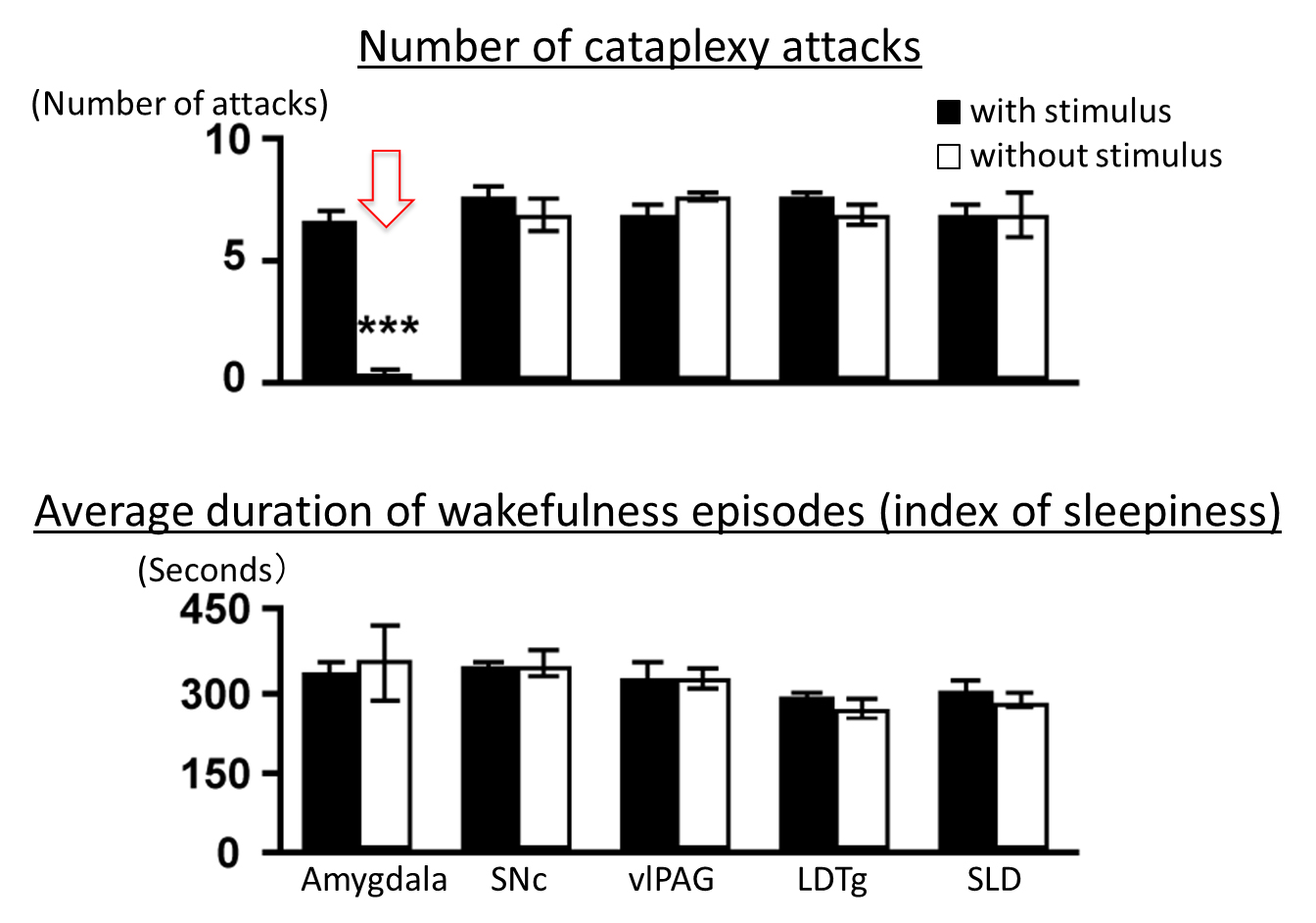
Serotonin neurons in the dorsal raphe nucleus extend projections throughout the brain and send information. In this study, with an optogenetic*6 tool, the team has discovered that catalepsy was almost completely inhibited by artificial augmentation of serotonin release induced by selectively stimulating serotonin nerve terminals in the amygdala in the narcolepsy model mice*7. The same experimental operation in the other brain region that controls REM sleep did not inhibit cataplexy. In addition, the team found that serotonin release reduced the amygdala activity. When the amygdala activity was artificially reduced in a direct manner, cataplexy was inhibited, while artificially augmented, frequency of cataplexy attack increased. Furthermore, the effect of orexin neurons inhibiting cataplexy was found to be abolished when serotonin release was inhibited selectively in the amygdala.
[Significance]
Cataplexy takes place, triggered by a sudden emotional excitement of positive valence such as a big laughter. This study has revealed that serotonin neurons do not directly suppress muscle tone weakening but inhibit cataplexy by reducing and controlling activities of the amygdala, which is involved in communicating emotional excitement. In fact, it is known that the amygdala of narcolepsy patients without orexin neurons responds excessively when the patients see, for example, interesting photos. By identifying neuronal pathway, orexin neuron – serotonin neuron in the dorsal raphe nucleus – the amygdala, the team believes that the current study has made a big step forward to understanding of the whole picture of the narcolepsy mechanism. It is also highly expected that new therapy would be developed for cataplexy.

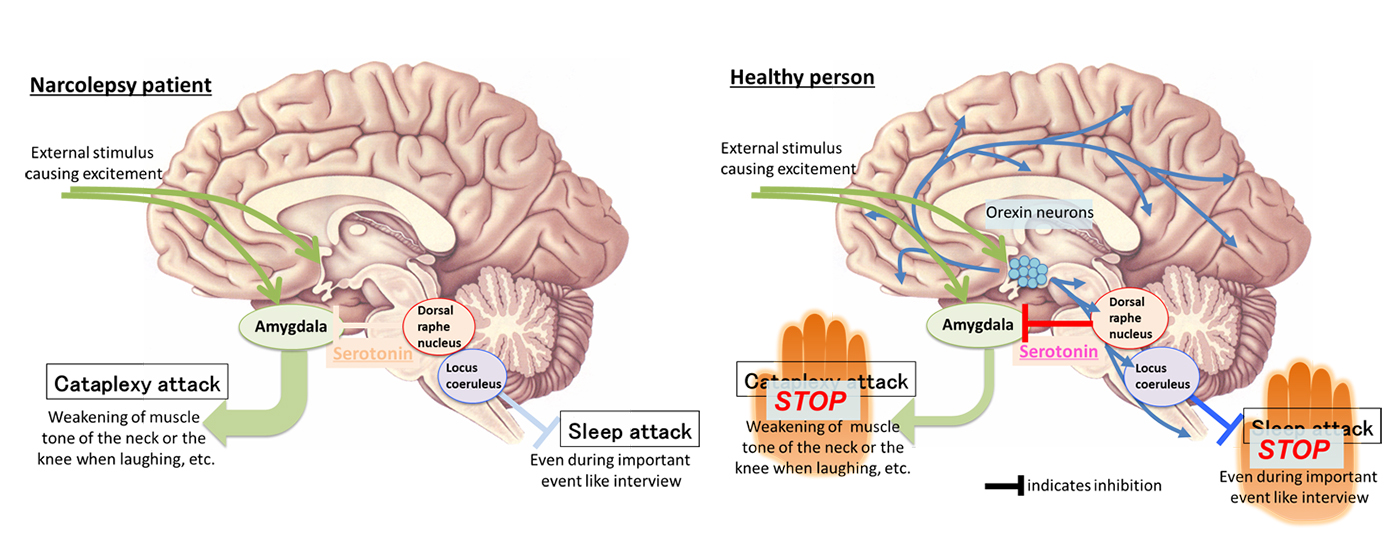
Figure 1. Cataplexy-inhibiting neuronal pathway revealed in this study.
External stimulus causing excitement such as laughter by a joke augments the amygdala activity. In narcolepsy patient (left) lacking orexin neurons, activities of the amygdala become excessive, causing cataplexy. In healthy person (right), orexin neurons augment the activities of serotonin neurons in the dorsal raphe nucleus, which reduce activities of the amygdala due to increased release of serotonin in the amygdala, which in turn inhibits cataplexy.
 Figure 2. Stimulation of serotonin nerve terminals in the amygdala inhibits cataplexy.
When serotonin nerve terminals in the amygdala are stimulated to augment serotonin release, cataplexy is inhibited (indicated by arrow in the upper part) while sleepiness is not inhibited (lower part). On the other hand, stimulation of serotonin nerve terminals in the other region controlling REM sleep does not induce any changes. LDTg, laterodorsal tegmental nucleus; SLD, sublaterodorsal nucleus; SNc, substantia nigra compact part; vlPAG, ventrolateral periaqueductal gray.
[Glossary]
*1 Orexin
Orexin A and orexin B are neuropeptides produced from a single gene in certain neurons of the hypothalamus. They consist of about 30 amino acid residues and function as neurotransmitters to convey information between neurons. Orexin producing neurons (orexin neurons) extend nerve projections throughout the brain. Orexin released from the orexin nerve terminal exerts different functions in various regions of the brain. It is known to prevent narcolepsy by stably maintaining wakefulness as well as to function for promotion of eating and metabolism and of response for reward.
*2 Narcolepsy
A sleep disorder characterized with excessive daytime sleepiness and with cataplexy. Caused by degeneration and loss of orexin neurons. Most patients experience their first narcolepsy symptoms in adolescence, and it is said that one patient is found out of 500 to 2000 persons.
*3 Cataplexy
Cataplexy is triggered by strong emotion and marked by sudden weakening of muscle tone of the whole body, the knee, the low back, the jaw, or the eyelid, but without consciousness impairment in many cases. One of the major characteristics of narcolepsy.
*4 Serotonin
Serotonin is one of the physiologically active amines and functions as a neurotransmitter to convey information between neurons in the brain. Serotonin producing neurons are found in specific regions of the brain and the dorsal raphe nucleus is one such region. Serotonin neurons extend projections throughout the brain. Serotonin released from the nerve terminals is involved in a wide variety of brain functions. Since the activities of serotonin neurons are high during wakefulness but low during sleep, serotonin is thought to be involved in regulating wakefulness and sleep. There is a hypothesis that low level of serotonin in the brain is one of the causes of depression.
*5 Amygdala
A brain region playing essential roles in processing emotional responses as well as in emotional memory. Emotion is defined as a temporal and big change of feelings induced rather in an acute manner, such as anger, terror, delight and sorrow. Emotion is accompanied with physical, physiological, and behavioral changes, and is distinguished from mood, that signifies weak feelings prevailing for a mid- and long-term in a mild manner.
*6 Optogenetics
Technique to manipulate the functions of cells by exogenously expressing light-activatable proteins and illuminating light.
*7 Narcolepsy model mouse
Genetically modified mouse, which lacks signal transduction by orexins, such as orexin-gene knockout mouse and orexin-receptor-gene knockout mouse. Narcolepsy model mouse exhibits symptoms similar to those of a narcolepsy patient. In this study, genetically modified mice with their orexin neurons being degenerated are used as narcolepsy model mice.
Article
Title: Serotonin neurons in the dorsal raphe mediate the anticataplectic action of orexin neurons by reducing amygdala activity
Journal: Proceedings of the National Academy of Sciences of the United States of America (2017)
Authors: Emi HASEGAWA1, Takashi MAEJIMA1, Takayuki YOSHIDA2, Olivia A. MASSECK3, Stefan HERLITZE3, Mitsuhiro YOSHIOKIA2, Takeshi SAKURAI1, Michihiro MIEDA1
1Kanazawa University, Japan; 2Hokkaido University, Japan; 3Ruhr-University Bochum, Germany
Doi: 10.1073/pnas.1614552114
Funder
JSPS KAKENHI, Astellas Foundation, Brain Science Foundation, Deutsche Forschungsgemeinschaft.


 PAGE TOP